muscle and tendon
extensor digitorum longus
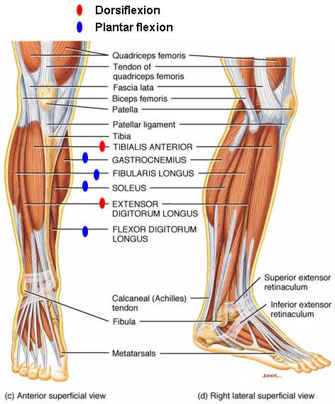
The extensor digitorum longus muscle is situated along the outside of the lower leg, just behind the tibialis anterior. It comes from close to the midline of the tibia and the shaft of the fibula. Its tendon divides into four parts as it passes over the front of the ankle. These parts continue over the surface of the foot and attach to the four smaller toes This muscle extends the second through fifth toes, and it dorsiflexes the foot through the ankle joint. Extension of the toes involves raising the toes up. Dorsiflexion of the foot involves flexing or raising the foot by bending the ankle joint up.
Learn More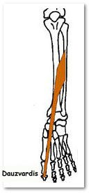
extensor hallucis longus
ORIGIN Middle half of anterior shaft of fibula INSERTION Base of distal phalanx of great toe ACTION Extends big toe and foot. Inverts foot and tightens subtalar joints NERVE Deep peroneal nerve
Learn Moretibialis anterior
Origin: Lateral condyle of tibia, proximal 1/2 - 2/3 or lateral surface of tibial shaft, interosseous membrane, and the deep surface of the fascia cruris Insertion: Medial and plantar surfaces of 1st cuneiform and on base of first metatarsal Action: Dorsiflexor of ankle and invertor of foot
Learn More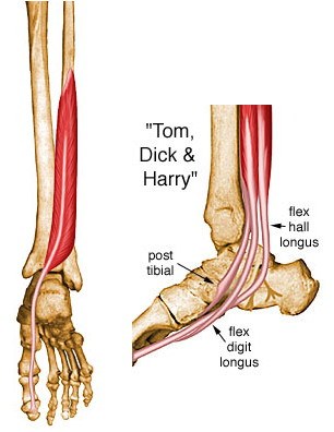
flexor hallucis longus
The flexor hallucis longus muscle is one of the three major deep muscles found in the lower back region of the leg. Specifically, the muscle spans part of the calf. It is the largest and strongest deep muscle of the leg's posterior section.
Learn More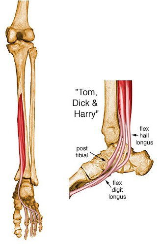
flexor digitorum longus
ORIGIN Posterior shaft of tibia below soleal line and by broad aponeurosis from fibula INSERTION Base of distal phalanges of lateral four toes ACTION Flexes distal phalanges of lateral four toes and foot at ankle. Supports lateral longitudinal arch NERVE Tibial nerve (S1, 2)
Learn More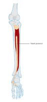
tibialis posterior
The tibialis posterior muscle is a relatively small, centrally located muscle present on the back side of the leg. This muscle is located between the two bones fibula and tibia in the lower leg and descends down to connect with the various other bones through the ankle. Insertion: (distal attachments): Navicular tuberosity, cuneiforms, cuboid, 2-4 metatarsals, and sustentaculum tali of calcaneus.
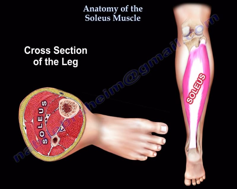
soleus
The soleus is the plantar flexor muscle of the ankle. It is capable of exerting powerful forces onto the ankle joint. It is located on the back of the lower leg and originates at the posterior (rear) aspect of the fibular head and the medial border of the tibial shaft. Origin: Laterally to the the head and the superior 1/3 of the fibula Medially to the middle 1/3 of the medial border of the tibia and the interosseous membrane Insertion: Into the calcaneus via the Achilles tendon Actions: Plantarflexion of the foot at the ankle Innervation: Tibial nerve (S1, S2 ) Blood Supply: Branches from the popliteal and posterior tibial arteries
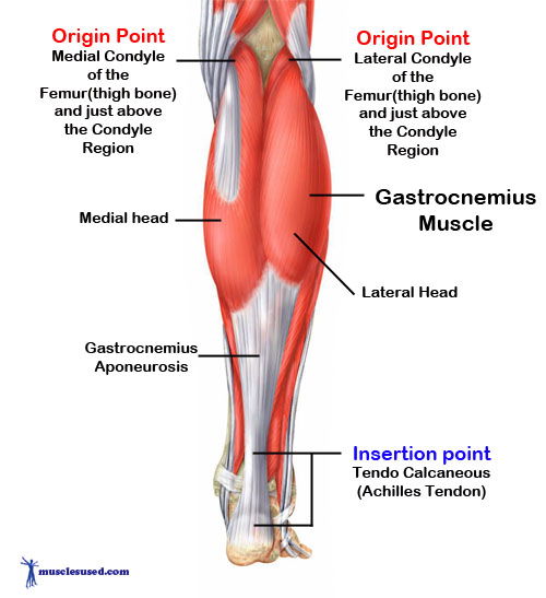
gastrocnemius
Plantarflexion is a movement required in jumping, running and walking. The gastrocnemius is a large muscle in the posterior compartment of the leg, and is the powerful muscle that enables plantarflexion, as well as knee flexion. In this article we will discuss the anatomy of the gastrocnemius muscle, together with the clinical relevance of this muscle.
peroneus brevis
Also known as the peroneus brevis, the fibularis brevis is a short, peroneal muscle that lies just underneath the peroneus longus muscle. The peroneal muscles extend along the outer portion of the lower leg and foot. The peroneus brevis attaches to the lower two-thirds of the fibula bone and the fifth metatarsal bone of the foot. The superficial peroneal nerves (L5 and S1) are the power source behind this muscle.The peroneus brevis plays an important role in the motor functions of the foot. The muscle assists in the flexion that moves the ball of the foot away from the body.
peroneus longus
The peroneus longus muscle, whose name means “long muscle of the fibula” is a major muscle of the lower leg that plantar flexes and everts the foot at the ankle. Also known as the fibularis longus, it is the longest muscle that attaches to the fibula and is used specifically when balancing one’s weight on one foot. originates at the head and the upper body of the fibula and the intermuscular septa. It inserts at the plantar side of the medial cuneiform and first metatarsal bone.
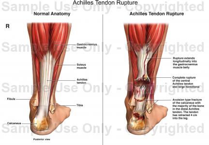
achilles tendon
The calcaneal tendon, also known as the tendon of Achilles, is a posterior leg tendon — a fibrous connective tissue that joins muscles in the back of the leg. It is formed when the soleus muscle tendon joins with the gastrocnemius tendon.The contracting calf muscles lift the heel by this tendon, thus producing a foot action that is basic to walking, running, and jumping. The Achilles tendon is the thickest and most powerful tendon in the body. If the tendon is cut, use of the leg for running or jumping is lost